Pixclara® (18F-FET) Can Help Pinpoint Glioma Biology1
Actor portrayals
18F-FET PET MOA1
- Fluorine-18 (F 18), the radiotracer used in Pixclara®, is a β+ emitting radionuclide that enables PET imaging
- 18F-FET is transported across the blood-brain barrier via LAT1
Amino acid transporters in the blood-brain barrier2
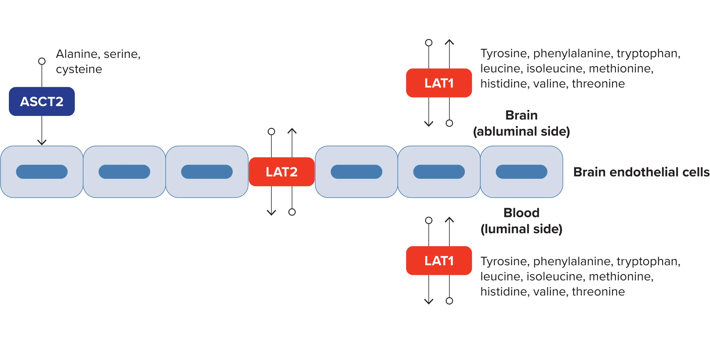
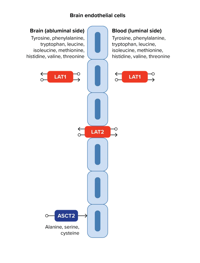
Adapted from Zaragozá R. Front Physiol. 2020;11:973. Under a CC BY Creative Commons Attribution License.
See how Pixclara® targets glioma
Molecularly targeted clarity: high-contrast, high-sensitivity brain tumor imaging1,3

Pixclara® is a radiolabeled derivative of tyrosine, an amino acid with high brain uptake within the brain lesion, even in the absence of blood-brain barrier disruption1,3

LAT transporters are highly active in glioma cells, leading to floretyrosine F 18 accumulation1

Accumulation within the tumor allows Pixclara® to provide clear imaging to differentiate recurrent or progressive glioma from treatment-related change, in conjunction with other diagnostic evaluations1,3

Pixclara® yields high TBR3

Pixclara® allows for high molecular sensitivity in tumor visualization3
Learn about reimbursement and coding for Pixclara®.
Get Information18F-FET, 18F-fluoro-ethyl-tyrosine; ASCT2, alanine-serine-cysteine transporter 2; LAT1, L-type (large neutral) amino acid transporter 1; LAT2, L-type (large neutral) amino acid transporter 2; MOA, mechanism of action; TBR, tumor-to-background ratio.