
When the Scan Is Unclear, so Is the Plan
Actor portrayal
Inconclusive differentiation between recurrence, progression, and treatment-related change can undermine patient management1
MRI and commonly used PET tracers like FDG PET can produce inconclusive results1
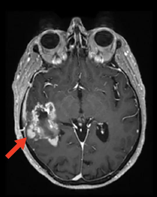
Limitations of CE MRI2
- May underestimate tumor size and growth because it nonspecifically reflects vascular pathology1,3,4
- Failure to accurately delineate tumor volumes can lead to undertreatment4
- Falsely detecting treatment-related change as tumor recurrence or progression can lead to overtreatment
Reprinted from Marner L, et al, under a Creative Commons 4.0 license. Clin Transl Imaging. 2017;5(2):135-149.
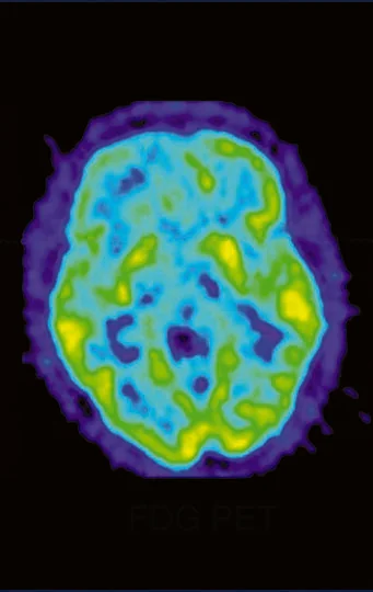
Limitations of FDG PET1
- Can be difficult to image due to high glucose metabolism in healthy brain tissue1
- Physiologic uptake in the brain parenchyma results in poor TBR5
Galldiks N, Kaufmann TJ, Vollmuth P, et al. Challenges, limitations, and pitfalls of PET and advanced MRI in patients with brain tumors: a report of the PET/RANO group. Neuro Oncol. 2024;26(7):1181-1194, by permission of Oxford University Press on behalf of the Society for Neuro-Oncology.
Commonly used diagnostics may mischaracterize tumors.
Learn MoreCE, contrast-enhanced; FDG, fluorodeoxyglucose-18; MRI, magnetic resonance imaging; PET, positron emission tomography; RANO, Response Assessment in Neuro-Oncology; TBR, tumor-to-background ratio.