Pixclara® (18F-FET) Helped Improve Clinical Decision-Making1
Actor portrayals
Real patient case 1: Tumor recurrence vs treatment-related change1
Recurrent glioblastoma, IDH-wildtype, post subtotal resection and chemoradiation
Patient background
- Male in his 50s with IDH-wildtype, MGMT-unmethylated glioblastoma
- CNS WHO grade 4
- Subtotal resection + chemoradiation + 5 cycles of adjuvant temozolomide
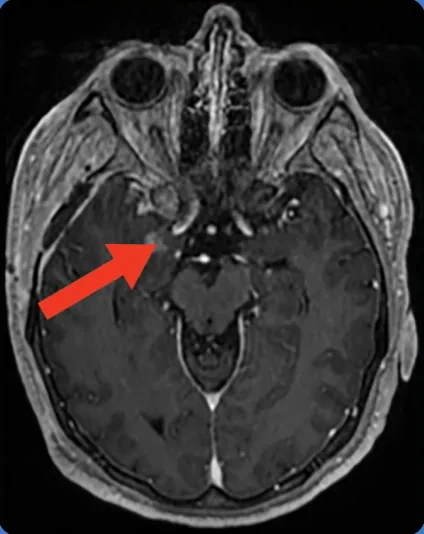
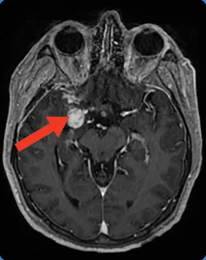
- 8 months later MRI showed an enlarging enhancing lesion with edema, concerning for recurrence vs radiation necrosis
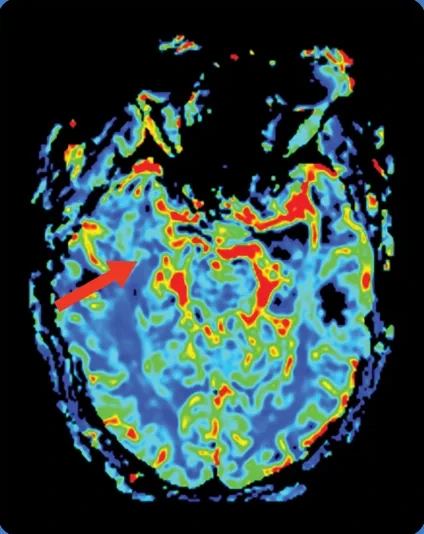
Interpretation of MRI: Lesion was assessed as indeterminate due to discrepancies between the results of conventional imaging and advanced MRI
Pixclara® detected: Residual viable disease (confirmed with biopsy)
Impact on clinical decision-making: PET metabolic activity confirmed viable residual disease, not radiation change. Surgeon proceeded with lesion excision
How did Pixclara® help clinicians?
- MRI alone could not distinguish recurrence from pseudoprogression
- Pixclara® clarified the MRI findings by indicating tumor progression vs radiation necrosis. Surgeon proceeded with re-resection
MRI follow-up
5 months before PET
(arrow showing enhancing lesion)
Conventional imaging
MRI follow-up
1.5 months before PET
(arrow showing enhancing lesion)
Conventional imaging
rCBV perfusion2
MRI follow-up
(arrow showing enhancing lesion)
PET: Lesion TBRmax = 4.66
TBRmean = 2.14
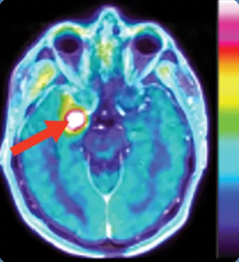
Scan with Pixclara®
confirming recurrent tumor
Real patient case 2: Tumor recurrence vs treatment-related change1
Recurrent glioblastoma, IDH-wildtype, post gross total resection, chemoradiation, and adjuvant temozolomide
Patient background
- Male in his 70s with glioblastoma, MGMT-unmethylated
- CNS WHO grade 4
- Gross total resection 13 months earlier
- Radiation completed 12 months earlier with temozolomide
- Adjuvant temozolomide completed 6 months earlier
- MRI raised concern for recurrent disease
Interpretation of MRI: Right parietal lesion concerning for recurrent glioblastoma vs treatment-related change before amino acid PET
Pixclara® detected: Residual viable disease, not radiation change (confirmed by biopsy)
Impact on clinical decision-making: Surgeon proceeded to lesion excision
How did Pixclara® help clinicians?
- MRI enhancement alone could not resolve what part of the lesion represented active tumor vs treatment effect
- Pixclara® enabled intralesional assessment based on differential uptake, highlighting a small focus suspicious for recurrence and separating it from adjacent treatment-related changes
- Surgeon proceeded with resection 3 weeks after PET/MRI
MRI follow-up
3 months before PET
(arrow showing enhancing lesion)
Conventional imaging
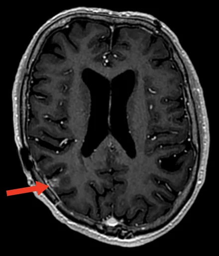
MRI follow-up
1 month before PET
(arrow showing enhancing lesion)
Conventional imaging
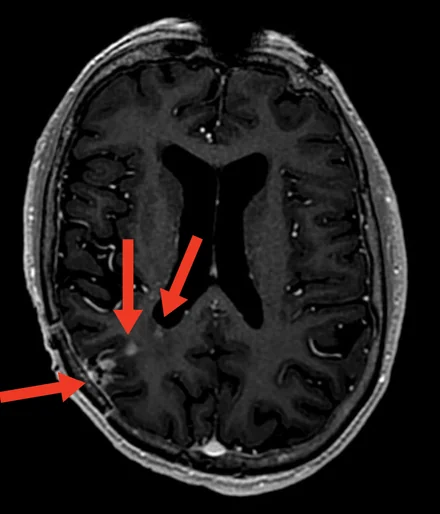
rCBV perfusion2
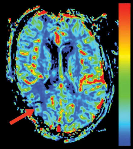
MRI follow-up
(arrow showing enhancing lesion)
PET: Lesion TBRmax = 3.23
TBRmean = 2.03
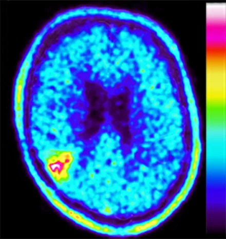
Pixclara® scan
confirming tumor
Real patient case 3: Tumor recurrence vs post-hemorrhagic or treatment-related change1
Recurrent astrocytoma, IDH-mutant, post subtotal resection and adjuvant vorasidenib
Patient background
- Female in her 20s with astrocytoma, IDH-mutant, MGMT-unmethylated
- CNS WHO grade 3
- Subtotal resection 23 months earlier
- Slow progression observed 11 months earlier; vorasidenib initiated
- Spontaneous intracranial hemorrhage 9 months ago
Interpretation of MRI: Lesion concerning for recurrent disease vs post-hemorrhagic or treatment-related change, with an evolving appearance across serial scans
Pixclara® detected: Distinct residual/recurrent astrocytoma
Impact on clinical decision-making: Surgeon proceeded to lesion excision
How did Pixclara® help clinicians?
- MRI alone could not distinguish what area of the evolving lesion represented active tumor vs post-hemorrhagic or treatment-related change
- Pixclara® enabled lesion assessment based on differential 18F-FET uptake, identifying residual/recurrent astrocytoma
- Surgeon proceeded with resection following imaging results
MRI follow-up
5 months before PET
(arrow showing enhancing lesion)
Conventional imaging
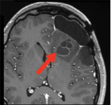
MRI follow-up
16 days before PET
(arrow showing enhancing lesion)
Conventional imaging
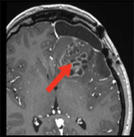
MRI follow-up
(arrow showing enhancing lesion)
PET: Lesion TBRmax = 3.10
TBRmean = 2.15
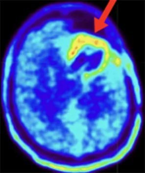
Scan with Pixclara®
confirming recurrent tumor
NCCN Clinical Practice Guidelines in Oncology (NCCN Guidelines®) Evidence NCCN Category 2A recommend 18F-FET PET imaging.3
Learn More18F-FET, 18F-fluoro-ethyl-tyrosine; CNS, central nervous system; IDH, isocitrate dehydrogenase; MGMT, O6-methylguanine-DNA methyltransferase; MRI, magnetic resonance imaging; NCCN, National Comprehensive Cancer Network; PET, positron emission tomography; rCBV, relative cerebral blood volume; TBRmax, maximum tumor-to-background ratio; TBRmean, mean tumor-to-background ratio; WHO, World Health Organization.